Muscular System: Power in Motion, Listing all 600 muscles in the human body
The muscular system is the human body‘s powerhouse, driving movement, providing stability, and enabling actions ranging from the subtle to the powerful. Comprising various types of muscles, each with distinct functions, this system is fundamental to our ability to interact with the world around us.
Overview of the Muscular System
The muscular system is a complex network of tissues that contract and relax, generating force and movement. This system includes over 600 muscles, varying in size and function, and is responsible for both voluntary and involuntary movements. From the blink of an eye to a hearty laugh, every action involves the coordination of muscles.
Listing all 600 muscles in the human body
Listing all 600 muscles in the human body individually would be an extensive and exhaustive task. Instead, I can provide you with a categorized overview of the major muscle groups. Keep in mind that this is a general classification, and there are more muscles within each group. Here are the major muscle groups in the human body:
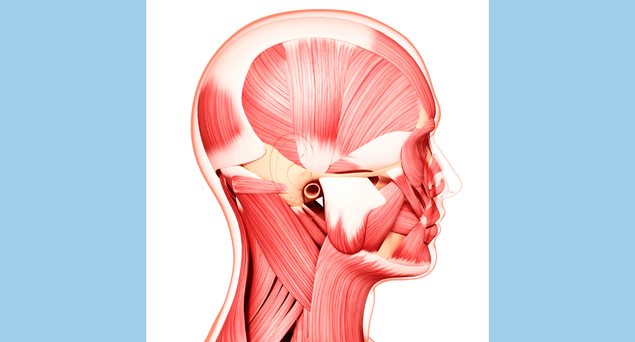
Head and Neck Muscles:
- 1. Frontalis
- 2. Temporalis
- 3. Masseter
- 4. Sternocleidomastoid
- 5. Trapezius
Shoulder Muscles:
- 6. Deltoid
- 7. Supraspinatus
- 8. Infraspinatus
- 9. Teres major
- 10. Subscapularis
Arm Muscles:
- 11. Biceps brachii
- 12. Triceps brachii
- 13. Brachialis
- 14. Brachioradialis
Chest Muscles:
- 15. Pectoralis major
- 16. Pectoralis minor
- 17. Serratus anterior
Abdominal Muscles:
- 18. Rectus abdominis
- 19. External oblique
- 20. Internal oblique
- 21. Transversus abdominis
Back Muscles:
- 22. Latissimus dorsi
- 23. Erector spinae group (spinalis, longissimus, iliocostalis)
- 24. Rhomboids
- 25. Levator scapulae
Pelvic Muscles:
- 26. Gluteus maximus
- 27. Gluteus medius
- 28. Gluteus minimus
- 29. Piriformis
Thigh Muscles:
- 30. Quadriceps femoris group (rectus femoris, vastus lateralis, vastus medialis, vastus intermedius)
- 31. Hamstring group (biceps femoris, semitendinosus, semimembranosus)
- 32. Adductor group (adductor magnus, adductor longus, adductor brevis)
Leg Muscles:
- 33. Gastrocnemius
- 34. Soleus
- 35. Tibialis anterior
- 36. Peroneus longus
Hand Muscles:
- 37. Intrinsic muscles of the hand (interossei, lumbricals, thenar, hypothenar muscles)
There are many more smaller muscles throughout the body that contribute to various movements and functions. This list covers the major muscle groups, and each group contains multiple individual muscles.
Remember, this is a simplified overview, and the human body is incredibly complex, with numerous smaller muscles contributing to overall function and movement.
Head and Neck Muscles:
Absolutely, let’s delve into more details about the head and neck muscles you’ve listed:
1. Frontalis:
Location: Located in the forehead.
Function: Mainly responsible for raising the eyebrows and creating wrinkles on the forehead.
2. Temporalis:
Location: Situated on the sides of the skull above the ears.
Function: Primarily involved in jaw movement, particularly in closing the jaw during chewing.
3. Masseter:
Location: Covers the sides of the jaw.
Function: One of the main muscles used for chewing; it elevates the mandible (lower jaw) during biting and chewing.
4. Sternocleidomastoid:
Location: Runs along each side of the neck.
Function: Allows for rotation and flexion of the head. When both muscles work together, they flex the neck; individually, they turn the head to the opposite side.
5. Trapezius:
Location: Extends down the back of the neck and upper spine.
Function: It has various functions, including supporting the shoulder blades, rotating and turning the head, and moving the shoulders.
These muscles collectively play crucial roles in facial expressions, mastication (chewing), head movements, and maintaining the structural integrity of the neck and upper back. The coordination of these muscles allows for a wide range of movements and functions, emphasizing the intricate design of the human musculature.
Shoulder Muscles:
Certainly, let’s explore the shoulder muscles you’ve listed in more detail:
6. Deltoid:
Location: Covers the shoulder joint.
Function: The deltoid is responsible for the abduction, flexion, and extension of the arm. It gives the shoulder its rounded shape and is involved in various arm movements.
7. Supraspinatus:
Location: Situated on the top of the shoulder blade.
Function: Initiates the abduction of the arm and stabilizes the shoulder joint during arm movement.
8. Infraspinatus:
Location: Occupies the lower part of the shoulder blade.
Function: Primarily responsible for the external rotation of the arm and stabilization of the shoulder joint.
9. Teres Major:
Location: Lies underneath the teres minor.
Function: Assists in the extension, adduction, and internal rotation of the arm. It works in conjunction with the latissimus dorsi.
10. Subscapularis:
Location: Positioned on the anterior surface of the scapula.
Function: The subscapularis is the primary muscle involved in the internal rotation of the arm and contributes to the stability of the shoulder joint.
These shoulder muscles collectively play a crucial role in providing both stability and a wide range of motion to the shoulder joint. They are integral for various arm movements, from lifting and rotating to reaching and throwing.
Arm Muscles:
Certainly, let’s dive into the details of the arm muscles you’ve listed:
11. Biceps Brachii:
Location: Located in the front of the upper arm.
Function: The biceps brachii is responsible for flexing the elbow joint, supinating the forearm (rotating the palm upward), and assisting in shoulder flexion.
12. Triceps Brachii:
Location: Situated on the back of the upper arm.
Function: The triceps brachii is the primary extensor of the elbow joint. It straightens the arm and is crucial for actions such as pushing and lifting.
13. Brachialis:
Location: Positioned underneath the biceps brachii.
Function: The brachialis is a powerful elbow flexor, assisting in bending the arm at the elbow joint. It is especially active during activities like lifting.
14. Brachioradialis:
Location: Found in the forearm, along the radius bone.
Function: The brachioradialis assists in flexing the forearm at the elbow. It is particularly active during activities that involve a hammering or pronated (palmdown) grip.
These arm muscles work in coordination to facilitate a variety of movements, including lifting, pushing, and bending at the elbow joint. Their specific functions contribute to the versatility of arm motion and play important roles in everyday activities.
Chest Muscles:
Certainly! Let’s explore the chest muscles you’ve listed:
15. Pectoralis Major:
Location: Large muscle covering the upper chest.
Function: The pectoralis major is responsible for the flexion, adduction, and medial rotation of the arm. It plays a crucial role in movements like hugging and bringing the arms across the chest.
16. Pectoralis Minor:
Location: Situated underneath the pectoralis major.
Function: The pectoralis minor assists in stabilizing the scapula (shoulder blade) and is involved in movements such as forward reaching and pulling the shoulder blades downward.
17. Serratus Anterior:
Location: Located on the lateral aspect of the ribcage.
Function: The serratus anterior plays a key role in the protraction and stabilization of the scapula. It is engaged in activities such as reaching forward, pushing, and raising the arms overhead.
These chest muscles contribute to the complex movements of the shoulder girdle, allowing for a wide range of arm motions. The pectoralis major, in particular, is a powerful muscle involved in various upper body exercises and functional activities.
Abdominal Muscles:
Certainly! Let’s delve into the details of the abdominal muscles you’ve listed:
18. Rectus Abdominis:
Location: Extends vertically along the front of the abdomen, commonly known as the “six-pack.”
Function: The rectus abdominis is responsible for flexing the spine, aiding in actions like situps. It also contributes to the stabilization of the pelvis.
19. External Oblique:
Location: Located on the sides and front of the abdomen, forming an angle.
Function: The external oblique muscles assist in the rotation and lateral flexion of the trunk. They also aid in flexing the spine forward.
20. Internal Oblique:
Location: Situated underneath the external obliques, running in the opposite direction.
Function: The internal oblique muscles work in conjunction with the external obliques, contributing to trunk rotation and lateral flexion. They also help stabilize the spine.
21. Trans-versus Abdominis:
Location: Deepest of the abdominal muscles, wrapping horizontally around the abdomen.
Function: The transversus abdominis provides stability to the spine and pelvis. It acts as a deep corset, aiding in abdominal compression and supporting internal organs.
These abdominal muscles play a vital role in core strength, providing support for various movements and helping maintain posture. The coordinated action of these muscles is crucial for activities such as bending, twisting, and maintaining a stable midsection.
Back Muscles:
Certainly! Let’s explore the back muscles you’ve listed:
22. Latissimus Dorsi:
Location: Broad muscle spanning the lower and middle back.
Function: The latissimus dorsi, often referred to as “lats,” is responsible for the adduction, extension, and medial rotation of the arm. It plays a significant role in movements like pulling, rowing, and arm extension.
23. Erector Spinae Group (Spinalis, Longissimus, Iliocostalis):
Location: These muscles run along the spine and are divided into three parts: spinalis, longissimus, and iliocostalis.
Function: The erector spinae group collectively plays a major role in spinal extension, lateral flexion, and rotation. They contribute to maintaining an upright posture and controlling movements of the vertebral column.
24. Rhomboids:
Location: Located between the shoulder blades.
Function: The rhomboids, including rhomboid major and rhomboid minor, retract and stabilize the scapula (shoulder blades). They are engaged in activities involving pulling the shoulder blades together.
25. Levator Scapulae:
Location: Positioned on the side and back of the neck.
Function: The levator scapulae elevates the scapula, assisting in movements like shrugging the shoulders. It also contributes to the rotation and lateral flexion of the neck.
These back muscles collectively contribute to the stability and movement of the spine, shoulders, and neck. They are essential for maintaining proper posture, facilitating various upper body movements, and providing support during activities such as lifting and pulling.
Pelvic Muscles:
Certainly! Let’s explore the pelvic muscles you’ve listed:
26. Gluteus Maximus:
Location: Largest of the three gluteal muscles, covering the buttocks.
Function: The gluteus maximus is the primary extensor of the hip joint. It plays a crucial role in actions like standing up from a seated position, climbing, and running.
27. Gluteus Medius:
Location: Situated on the outer surface of the pelvis.
Function: The gluteus medius is responsible for hip abduction and internal rotation. It helps in stabilizing the pelvis during activities like walking and maintaining balance.
28. Gluteus Minimus:
Location: Positioned underneath the gluteus medius.
Function: The gluteus minimus assists in hip abduction and internal rotation. It works in conjunction with the gluteus medius to support pelvic stability.
29. Piriformis:
Location: Deep within the buttock region, beneath the gluteal muscles.
Function: The piriformis aids in external rotation and abduction of the hip. It is also involved in stabilizing the sacroiliac joint.
These pelvic muscles contribute to the stability, strength, and mobility of the hip joint and pelvis. They play essential roles in various activities, including walking, running, and maintaining an upright posture.
Thigh Muscles:
Certainly! Let’s delve into the details of the thigh muscles you’ve listed:
30. Quadriceps Femoris Group (Rectus Femoris, Vastus Lateralis, Vastus Medialis, Vastus Intermedius):
Rectus Femoris:
Location: Runs along the front of the thigh.
Function: The rectus femoris is a part of the quadriceps group and is responsible for knee extension and hip flexion.
Vastus Lateralis, Vastus Medialis, Vastus Intermedius:
Location: These muscles are located on the front of the thigh.
Function: The vastus muscles collectively contribute to knee extension, playing a crucial role in movements like standing up and walking.
31. Hamstring Group (Biceps Femoris, Semitendinosus, Semimembranosus):
Biceps Femoris:
Location: Located on the back of the thigh.
Function: The biceps femoris is a part of the hamstring group and is involved in knee flexion and hip extension.
Semitendinosus and Semimembranosus:
Location: Also situated on the back of the thigh.
Function: These muscles are part of the hamstrings and contribute to knee flexion, hip extension, and stabilization of the knee.
32. Adductor Group (Adductor Magnus, Adductor Longus, Adductor Brevis):
Adductor Magnus:
Location: Positioned on the inner thigh.
Function: The adductor magnus contributes to hip adduction and extension.
Adductor Longus and Adductor Brevis:
Location: Also located on the inner thigh.
Function: These muscles assist in hip adduction and stabilization of the hip joint.
These thigh muscles collectively play essential roles in various leg movements, including walking, running, and maintaining balance. The quadriceps and hamstrings are particularly crucial for activities involving the knee joint, while the adductor muscles contribute to movements involving the hip joint.
Leg Muscles:
Let’s explore the details of the leg muscles you’ve listed:
33. Gastrocnemius:
Location: The twoheaded muscle located at the back of the lower leg.
Function: The gastrocnemius is a powerful muscle involved in plantarflexion of the foot, enabling actions like pointing the toes downward and pushing off the ground during walking or running.
34. Soleus:
Location: Situated beneath the gastrocnemius, also at the back of the lower leg.
Function: The soleus works alongside the gastrocnemius in plantarflexion. It is particularly active during activities that require sustained or slow movements, such as standing.
35. Tibialis Anterior:
Location: Found at the front of the lower leg.
Function: The tibialis anterior is responsible for dorsiflexion of the foot, allowing the toes to be lifted upward. It plays a key role in walking and helps control the descent of the foot during activities like going downstairs.
36. Peroneus Longus:
Location: Located on the outer side of the lower leg.
Function: The peroneus longus is involved in plantarflexion and eversion of the foot. It helps stabilize the ankle joint during walking and supports balance.
These leg muscles collectively contribute to the intricate movements of the foot and ankle, essential for activities such as walking, running, and maintaining balance. The gastrocnemius and soleus form the prominent muscles of the calf, while the tibialis anterior and peroneus longus play crucial roles in controlling foot movements. If you have more specific questions or would like information on other muscle groups, feel free to ask!
Hand Muscles:
Certainly! Let’s explore the details of the hand muscles you’ve listed:
37. Intrinsic Muscles of the Hand:
Interossei Muscles:
Location: Located between the metacarpal bones of the hand.
Function: The interossei muscles are responsible for the abduction and adduction of the fingers. They play a crucial role in controlling finger movements and grip.
Lumbrical Muscles:
Location: Found in the palm of the hand, connecting the tendons of the flexor digitorum profundus.
Function: The lumbrical muscles assist in flexing the metacarpophalangeal joints while extending the interphalangeal joints. They contribute to fine motor control in the fingers.
Thenar Muscles:
Location: Located at the base of the thumb.
Function: The thenar muscles, including the abductor pollicis brevis, flexor pollicis brevis, opponens pollicis, and adductor pollicis, control thumb movements and contribute to gripping and pinching actions.
Hypothenar Muscles:
Location: Situated at the base of the little finger.
Function: The hypothenar muscles, including the abductor digiti minimi, flexor digiti minimi brevis, and opponens digiti minimi, control movements of the little finger and contribute to grip strength.
These intrinsic hand muscles are essential for precise and coordinated movements of the fingers and thumb. They allow for a wide range of hand functions, from grasping and holding objects to intricate tasks requiring dexterity and fine motor control.
Types of Muscles
Understanding the diversity of muscles within the body adds depth to our appreciation of the muscular system. There are three main types of muscles:
1. Skeletal Muscles
Location: Attached to bones by tendons.
Voluntary/Involuntary: Voluntary.
Function: Enable movement of the skeleton, including actions like walking and grasping objects.
2. Smooth Muscles
Location: Found in the walls of internal organs (e.g., digestive tract, blood vessels).
Voluntary/Involuntary: Involuntary.
Function: Facilitate involuntary movements of internal organs, such as peristalsis in the digestive system.
3. Cardiac Muscles
Location: Exclusive to the heart.
Voluntary/Involuntary: Involuntary.
Function: Responsible for pumping blood throughout the body, maintaining the circulatory system.
Importance in Movement and Stability
The muscular system plays a pivotal role in our ability to move and maintain stability. Here’s why it’s so crucial:
1. Movement
Skeletal Muscles: Provide the force needed for voluntary movements, allowing us to walk, run, lift objects, and perform various activities.
Smooth Muscles: Facilitate involuntary movements in internal organs, ensuring processes like digestion and blood circulation.
Cardiac Muscles: Contract rhythmically to pump blood, maintaining circulation.
2. Stability
Skeletal Muscles: Act as stabilizers during movement, preventing joints from dislocating and maintaining posture.
Smooth Muscles: Contribute to the stability of internal organs by regulating the flow of substances within them.
Cardiac Muscles: Ensure the heart maintains a steady and efficient pumping rhythm, contributing to overall bodily stability.
The Harmonious Dance of Muscles
Picture the muscular system as a choreographed dance, where different types of muscles work together seamlessly. Skeletal muscles provide the strength for purposeful actions, while smooth muscles handle involuntary functions, and cardiac muscles maintain the steady beat that sustains life.
Understanding the intricacies of the muscular system not only enhances our appreciation for the body’s capabilities but also sheds light on the importance of maintaining muscle health. As we delve further into the wonders of human physiology, the muscular system stands as a testament to the incredible synergy within our bodies.
FAQs
1. Q: What is the Muscular System, and what does it consist of?
A: The Muscular System is a complex network of muscles that enable movement. It includes skeletal muscles, smooth muscles, and cardiac muscles.
2. Q: How do muscles contract, and what role does the nervous system play in muscle movement?
A: Muscle contraction is initiated by nerve impulses. The nervous system signals muscles to contract, generating the force necessary for movement.
3. Q: What are the different types of muscles in the Muscular System, and how do they function differently?
A: Skeletal muscles control voluntary movements, smooth muscles manage involuntary functions, and cardiac muscles specifically power the heart’s pumping action.
4. Q: Can exercise impact muscle health, and what role does it play in muscle development and maintenance?
A: Regular exercise promotes muscle strength and endurance. It stimulates muscle growth, prevents atrophy, and enhances overall muscular health.
https://nursingscholar101.com/human-body-systems-and-organs/